The Multiple Sclerosis Imaging Laboratory, directed by Caterina Mainero, MD, PhD, focuses on using advanced imaging modalities to investigate the heterogenous aspects of multiple sclerosis pathology that include neuroinflammation, demyelination, neurodegeneration, and tissue repair.
Our goal is to integrate novel imaging methods with clinical and biological markers of the disease to investigate the brain mechanisms underlying disease activity and progression, and to define the most sensitive neuroimaging tools for improving disease diagnosis and monitoring.
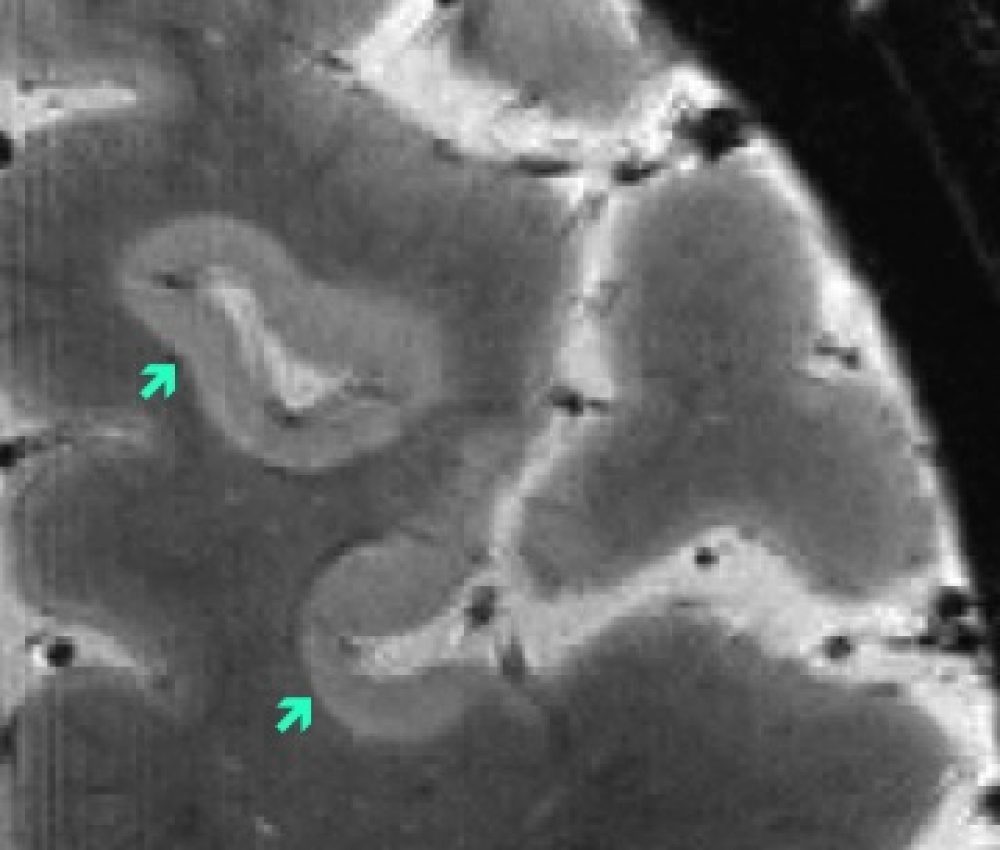
Cortical Pathology
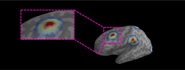
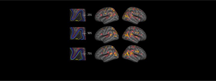
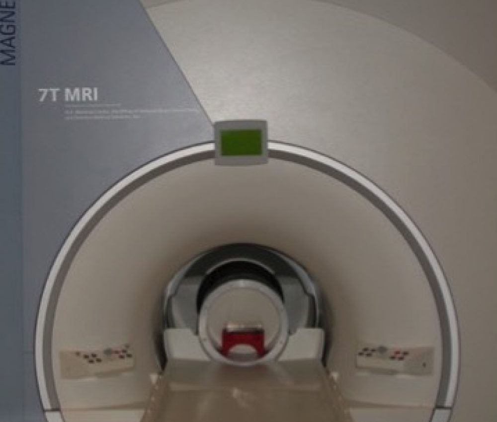
We use ultra high field 7 Tesla (7T) MRI to study lesions in the cortex of MS patients and their contribution to clinical outcome. We are developing quantitative imaging methods at 7T to investigate cortical neurodegeneration and tissue repair
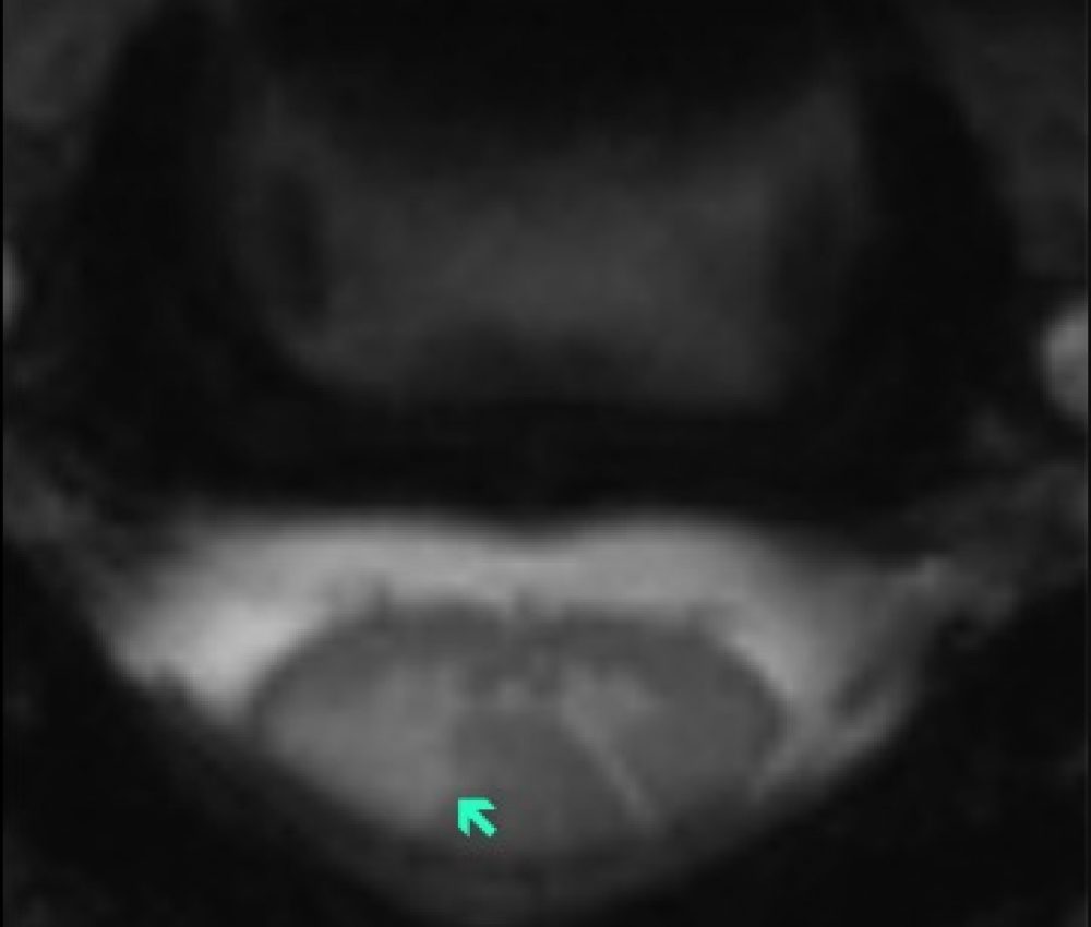
Spinal Cord Pathology
Spinal cord pathology is frequent in MS and can contribute to physical disability. Our lab is using 7T MRI to investigate the occurrence, distribution, and clinical impact of spinal cord pathology in both gray and white matter.
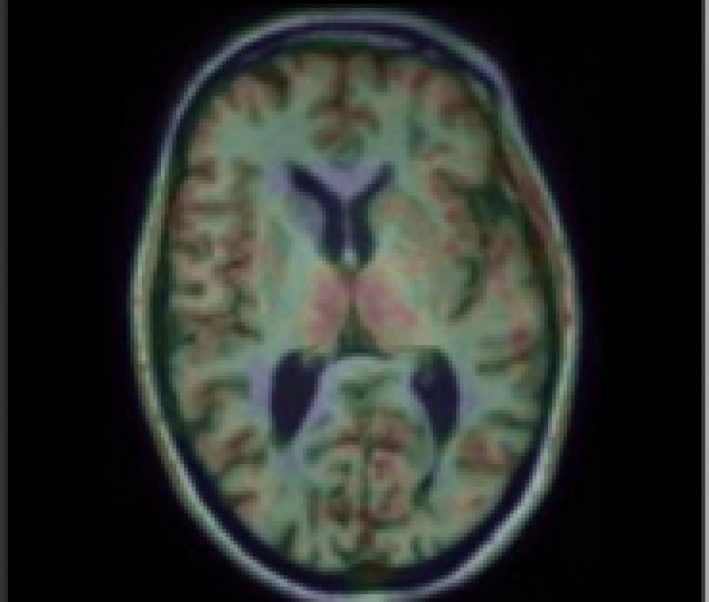
Neuroinflammation
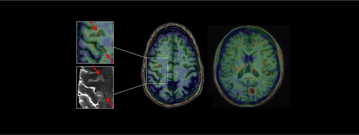
MR-PET imaging is used to study neuroinflammation in the brain of MS patients and assess its role in neurodegeneration and tissue repair, particularly in the cortex.
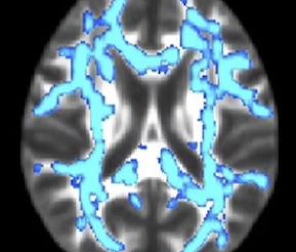
Axonal-Myelin Pathology
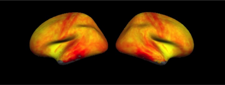
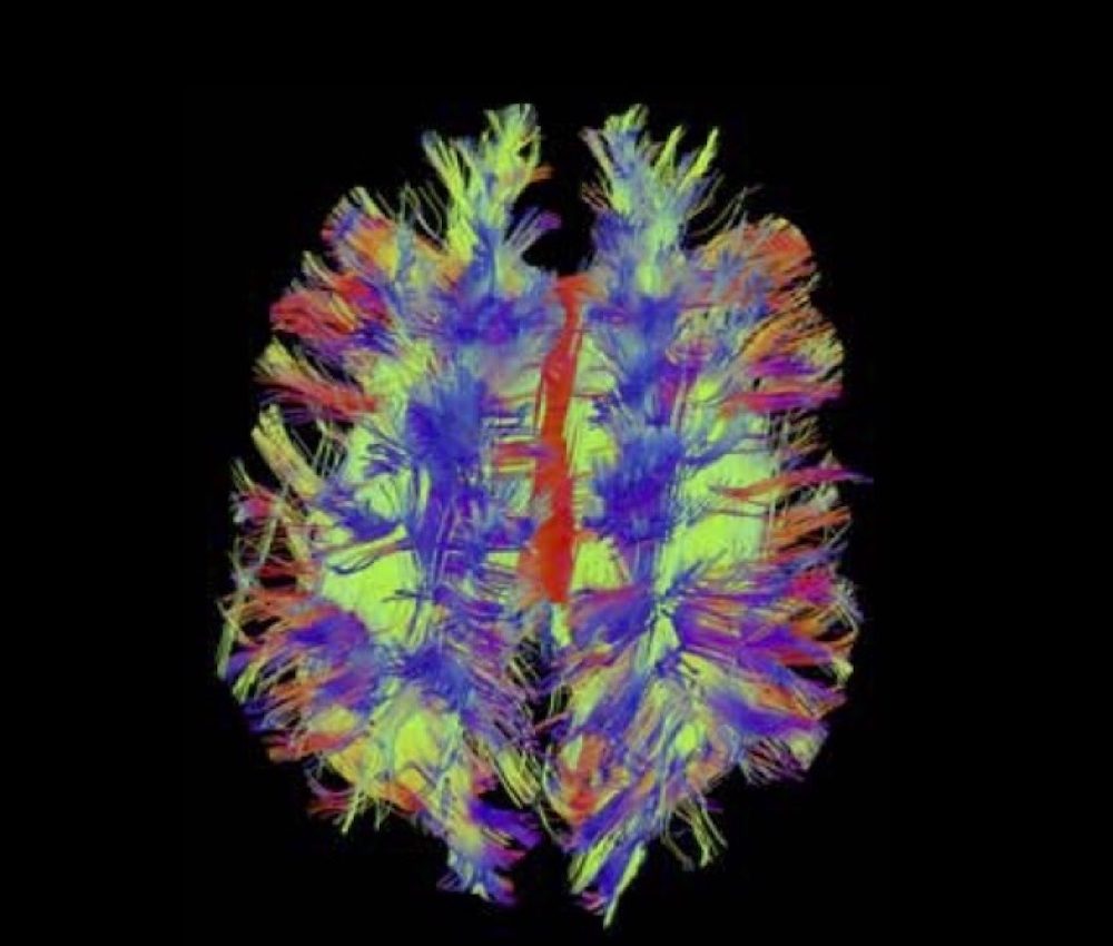
Axonal injury is a result of MS and a main determinant of neurological disability. With a dedicated 3T MRI scanner (Connectome), we can obtain advanced diffusion imaging protocols that can be used to measure changes in axonal and myelin integrity in vivo.
Brain Connectivity
Diffusion imaging can be used to assess structural brain connectivity in patients with MS, and how this may be affected by MS pathological processes that include demyelination, neurodegeneration and inflammation.
Clinical Trials
Our Lab is conducting a series of clinical studies to test the ability of novel imaging markers to characterize the heterogeneous pathology of MS in vivo and to predict disease outcome and/or response to therapies.

